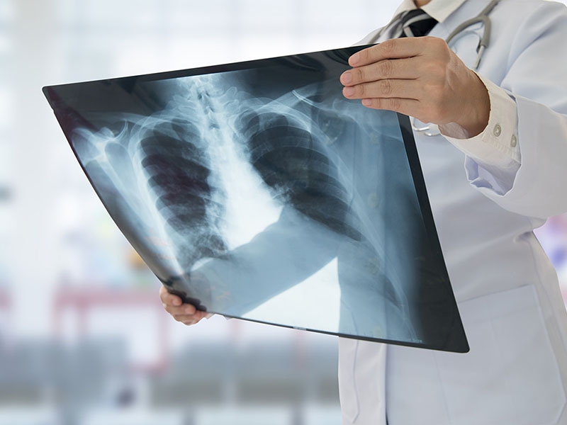
X-Ray
Watch our video about X-Ray
An X-ray is a non-invasive medical imaging technique used to visualize the internal structures of the body, including bones, organs, and tissues. This diagnostic test relies on electromagnetic radiation to create images that help detect injuries, infections, and various health conditions.
X-rays are among the most widely used imaging techniques in medicine, as they provide quick and detailed results with minimal discomfort for the patient. They are commonly used in orthopedics, dentistry, pulmonology, and emergency medicine to assess a wide range of medical conditions.
What is an X-Ray Used For?
X-rays serve multiple medical purposes, including:
- Diagnosing fractures, dislocations, and bone abnormalities.
- Detecting lung conditions such as pneumonia, tuberculosis, and lung cancer.
- Assessing dental issues, including cavities, impacted teeth, and jawbone infections.
- Identifying digestive system problems, such as bowel obstructions or swallowed foreign objects.
- Evaluating heart and vascular conditions, including enlarged heart or arterial blockages.
- Screening for tumors, infections, and abnormal masses in various parts of the body.
- Monitoring post-surgical recovery, ensuring proper healing of bones and implants.
Because X-rays are fast, safe, and widely available, they are a crucial tool for emergency diagnostics and routine health assessments.
How Does an X-Ray Work?
The X-ray procedure follows these steps:
- Patient Positioning: The patient is positioned according to the area being examined, either standing, sitting, or lying down.
- Radiation Exposure: A controlled X-ray beam is directed at the body, and the radiation passes through tissues to create an image. Dense structures (like bones) appear white, while soft tissues appear in shades of gray.
- Image Capture and Analysis: The images are recorded digitally or on film and analyzed by a radiologist or physician to detect abnormalities.
The process is painless and usually takes only a few minutes. In some cases, a contrast agent (such as barium or iodine) may be used to enhance the visibility of certain organs and blood vessels.
Types of X-Rays
There are several types of X-rays, each used for specific medical purposes. The most common types include:
1. Chest X-Ray
A chest X-ray is commonly used to evaluate lung and heart health. It helps detect conditions such as pneumonia, tuberculosis, lung cancer, and heart enlargement.
This type of X-ray is often ordered for patients experiencing chronic cough, chest pain, difficulty breathing, or suspected infections. Since it is quick and widely available, it is frequently used in emergency settings.
2. Bone X-Ray
A bone X-ray is used to examine fractures, joint dislocations, arthritis, and bone infections.
It provides a clear view of the skeletal system, allowing doctors to assess bone alignment, healing progress, and degenerative diseases. Orthopedic specialists commonly request bone X-rays for sports injuries, osteoporosis evaluations, and post-surgical follow-ups.
3. Abdominal X-Ray
An abdominal X-ray is performed to identify digestive system issues, such as bowel obstructions, kidney stones, or swallowed foreign objects.
In some cases, a contrast dye (barium swallow) is used to enhance visibility and assess problems in the stomach, intestines, and esophagus. Patients experiencing severe abdominal pain, bloating, or persistent digestive discomfort may require this exam.
4. Dental X-Ray
A dental X-ray is used to evaluate tooth and jaw health. It helps diagnose cavities, impacted teeth, infections, and bone loss in the jaw.
This type of X-ray is essential for orthodontic treatments, wisdom tooth extractions, and root canal planning. It is performed using low-radiation techniques, making it safe for routine dental checkups.
5. Fluoroscopy (Dynamic X-Ray Imaging)
Fluoroscopy is a specialized type of X-ray that provides real-time moving images of the body's internal functions.
It is commonly used for guiding catheter placements, barium swallow tests, and joint injections. This technique allows doctors to assess organ function dynamically, making it useful for gastrointestinal studies and cardiovascular exams.
What Conditions Can an X-Ray Detect?
X-rays help diagnose various medical conditions, including:
- Bone Fractures and Dislocations – Identifies breaks, cracks, and misaligned bones.
- Pneumonia and Lung Infections – Detects fluid accumulation and inflammation in the lungs.
- Osteoarthritis and Rheumatoid Arthritis – Reveals joint damage and cartilage deterioration.
- Heart Enlargement and Fluid Buildup – Assesses heart size and signs of heart failure.
- Kidney Stones and Gallstones – Identifies mineral deposits in the kidneys or gallbladder.
- Digestive Tract Blockages – Detects bowel obstructions, perforations, and foreign objects.
- Dental Abnormalities – Helps diagnose tooth decay, infections, and impacted teeth.
When is an X-Ray Recommended?
An X-ray is indicated in the following situations:
- After an Injury or Accident – To check for fractures, sprains, or internal damage.
- Persistent Chest Pain or Shortness of Breath – To rule out lung infections, heart problems, or rib fractures.
- Chronic Joint or Bone Pain – To assess arthritis, osteoporosis, or joint degeneration.
- Unexplained Digestive Symptoms – To detect intestinal obstructions, ulcers, or tumors.
- Suspected Tooth or Jaw Problems – For detecting cavities, misalignment, or infections.
- Screening for Tumors or Abnormal Growths – To check for cancer or benign masses.
- Pre-Surgical Evaluations – To assess the area before orthopedic, dental, or lung surgeries.
Pre and Post-X-Ray Care
Before the X-Ray:
- Remove metal objects, such as jewelry, as they may interfere with imaging.
- Inform the doctor if you are pregnant before undergoing the exam.
- Wear comfortable clothing, avoiding zippers or metal fastenings.
After the X-Ray:
- No recovery time is needed for standard X-rays.
- For contrast X-rays, drink plenty of fluids to help eliminate the contrast agent.
- Follow up with your doctor to discuss results and necessary treatments.
Contraindications for X-Ray
Although X-rays are generally safe, they may not be recommended for:
- Pregnant women, unless absolutely necessary.
- Patients with severe kidney disease, in cases requiring contrast dye.
- Young children, unless the benefits outweigh the risks.
In such cases, alternative imaging techniques may be recommended.
Alternatives for Patients Who Cannot Undergo an X-Ray
For individuals who cannot undergo an X-ray, alternative diagnostic tests include:
- MRI (Magnetic Resonance Imaging) – Provides detailed soft tissue imaging without radiation.
- CT Scan (Computed Tomography) – Offers high-resolution 3D images for in-depth diagnosis.
- Ultrasound – A radiation-free imaging option for pregnancy, soft tissue, and organ evaluations.
Schedule Your X-Ray at Clinic Consultation
X-ray services are available at Clinic Consultation, performed with modern imaging technology for accurate diagnosis. Whether you need an emergency scan, routine checkup, or injury assessment, our specialists ensure safe and high-quality medical imaging.
📅 Book your X-ray appointment today and receive professional care for your diagnostic needs!
Click here to schedule an appointment online