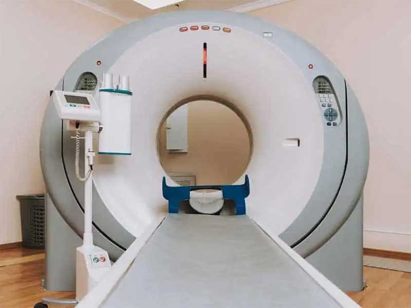
Magnetic Resonance Imaging (MRI)
Watch our video about Magnetic Resonance Imaging (MRI)
Magnetic Resonance Imaging (MRI) is a non-invasive imaging technique that uses magnetic fields and radio waves to create detailed images of internal organs, tissues, bones, and blood vessels. Unlike X-rays or CT scans, MRI does not use ionizing radiation, making it a safer option for repeated examinations.
This technology is particularly useful for diagnosing complex medical conditions, providing high-resolution images that help doctors assess neurological, musculoskeletal, and cardiovascular issues. MRI is widely used in various medical specialties, including neurology, orthopedics, oncology, and cardiology.
What is MRI Used For?
MRI is commonly used for diagnosing, monitoring, and planning treatments for various medical conditions, including:
- Detecting brain disorders, such as strokes, tumors, and multiple sclerosis.
- Evaluating spinal injuries and disc herniations in the vertebral column.
- Assessing joint problems, including torn ligaments, cartilage damage, and arthritis.
- Identifying cardiovascular conditions, such as heart defects and blood vessel abnormalities.
- Detecting tumors in soft tissues and organs like the liver, kidneys, and reproductive system.
- Examining the digestive system for issues such as Crohn’s disease and liver disease.
- Providing detailed imaging for surgical planning and treatment follow-up.
Due to its exceptional image clarity, MRI is a preferred method for complex diagnoses that require in-depth analysis.
How Does an MRI Work?
The MRI procedure follows these key steps:
- Patient Preparation: The patient is positioned on a movable table that slides into the MRI scanner. A contrast dye may be injected if detailed imaging is required.
- Magnetic Field Activation: The machine generates a strong magnetic field, which aligns hydrogen atoms in the body.
- Radio Waves and Image Capture: Radio waves are transmitted to the body, and the response signals are collected to form detailed, high-resolution images of internal structures.
The procedure is completely painless and typically lasts between 30 to 60 minutes, depending on the area being scanned. Patients must remain still during the process to ensure image accuracy.
Types of MRI
There are several types of MRI scans, each serving a specific purpose:
1. Brain MRI
A Brain MRI provides detailed images of the brain and spinal cord. It is used to diagnose neurological conditions such as brain tumors, multiple sclerosis, strokes, and aneurysms.
This scan is crucial for patients experiencing severe headaches, dizziness, seizures, or cognitive impairments. It helps neurologists detect abnormalities that may not be visible on other imaging tests.
2. Spine MRI
A Spine MRI is commonly used to evaluate back pain, nerve compression, and spinal cord injuries. It helps diagnose herniated discs, spinal tumors, and degenerative diseases affecting the vertebrae.
Unlike X-rays, which only show bones, MRI scans reveal soft tissues, nerves, and spinal discs, making it a preferred choice for detecting chronic pain conditions and post-surgical complications.
3. Musculoskeletal MRI
A Musculoskeletal MRI is used to examine joints, muscles, ligaments, and tendons. It is highly beneficial for sports injuries, arthritis, and post-trauma evaluations.
This scan provides precise imaging of ligament tears, cartilage damage, and inflammation, making it essential for orthopedic treatments and physical rehabilitation planning.
4. Cardiac MRI
A Cardiac MRI evaluates heart structure, function, and blood flow. It is used for diagnosing heart defects, myocardial infarctions (heart attacks), and cardiomyopathies.
This test provides detailed images of the heart's chambers, valves, and arteries, helping cardiologists assess heart health and plan treatments for cardiovascular diseases.
5. Abdominal and Pelvic MRI
An Abdominal MRI is used to examine organs such as the liver, kidneys, pancreas, and intestines. It is often requested for detecting tumors, cysts, liver disease, or inflammatory bowel disease (IBD).
A Pelvic MRI is used to assess reproductive organs, including the uterus, ovaries, and prostate, helping diagnose conditions like fibroids, ovarian cysts, and prostate cancer.
What Conditions Can MRI Detect?
MRI can help diagnose a variety of health conditions, including:
- Brain Tumors and Stroke – Detects abnormalities in brain tissue and blood flow.
- Spinal Cord Disorders – Identifies herniated discs, nerve compression, and spinal injuries.
- Arthritis and Joint Damage – Evaluates cartilage wear, inflammation, and ligament tears.
- Cancer Detection – Identifies tumors in soft tissues, organs, and bones.
- Heart and Blood Vessel Abnormalities – Assesses heart function and detects vascular diseases.
- Liver and Kidney Disease – Diagnoses liver cirrhosis, kidney cysts, and other organ issues.
- Pelvic Disorders – Helps diagnose fibroids, ovarian cysts, and prostate enlargement.
When is an MRI Recommended?
An MRI is recommended in various medical situations, including:
- Unexplained Headaches or Neurological Symptoms – To rule out brain tumors or nerve disorders.
- Persistent Back or Joint Pain – To assess musculoskeletal injuries and degenerative conditions.
- Cancer Screening and Tumor Evaluation – For detecting and monitoring abnormal growths.
- Unexplained Abdominal Pain – To check for organ inflammation, tumors, or gastrointestinal disorders.
- Heart Disease Diagnosis – For patients with suspected heart conditions.
- Pre-Surgical Planning – Provides detailed imaging before complex surgeries.
- Trauma or Injury Assessment – Helps identify internal bleeding, fractures, and soft tissue damage.
Pre and Post-MRI Care
Before the MRI:
- Remove metallic objects (jewelry, piercings, metal implants).
- Inform your doctor if you have claustrophobia, as sedation may be recommended.
- Avoid caffeine or stimulants, which may affect the results of brain MRIs.
After the MRI:
- No recovery time is needed, and normal activities can be resumed immediately.
- For contrast MRIs, drinking plenty of water helps flush out the contrast agent.
- Discuss results with your doctor to determine the next steps in your treatment plan.
Contraindications for MRI
MRI is generally safe, but it may not be recommended for individuals with:
- Metal implants, pacemakers, or defibrillators, as the magnetic field can interfere with these devices.
- Severe kidney disease, which may limit the use of contrast agents.
- Severe claustrophobia, unless sedation is administered.
For these cases, alternative imaging tests may be required.
Alternatives for Patients Who Cannot Undergo an MRI
For patients who cannot undergo an MRI, other imaging options include:
- CT Scan (Computed Tomography) – Offers detailed imaging using X-rays.
- Ultrasound – A radiation-free option for soft tissue evaluation.
- X-Ray – Effective for bone assessments and initial evaluations.
Schedule Your MRI at Clinic Consultation
MRI services are available at Clinic Consultation, performed with advanced imaging technology for accurate diagnoses. Whether you need an MRI for neurological, orthopedic, cardiac, or abdominal conditions, our specialists ensure safe and high-quality scanning.
📅 Book your MRI appointment today and receive expert medical imaging for your healthcare needs!
Click here to schedule an appointment online