Echocardiogram
Watch our video about Echocardiogram
An echocardiogram is a non-invasive imaging test that uses Ultrasound waves to create detailed images of the heart’s structure and function. It helps cardiologists assess how well the heart is pumping blood, detect abnormalities, and diagnose heart conditions.
This test is completely painless, does not use radiation, and can be performed on patients of all ages. An echocardiogram is one of the most essential tools in cardiology, allowing doctors to visualize the heart’s chambers, valves, blood flow, and muscle function in real time.
What is an Echocardiogram Used For?
An echocardiogram serves multiple medical purposes, including:
- Assessing overall heart function and detecting abnormalities.
- Diagnosing heart diseases, including congenital defects, valve issues, and heart failure.
- Monitoring heart conditions over time, especially after a heart attack.
- Checking for fluid accumulation around the heart (pericardial effusion).
- Evaluating heart valve function and identifying stenosis (narrowing) or regurgitation (leakage).
- Assessing blood flow and measuring pressure inside the heart chambers.
- Determining if a patient is fit for heart surgery or other interventions.
By providing real-time heart images, an echocardiogram is a crucial tool in diagnosing and managing cardiovascular health.
How Does an Echocardiogram Work?
The echocardiogram procedure follows these steps:
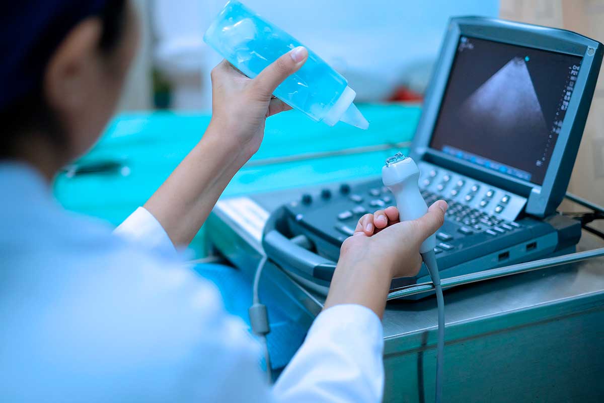
- Preparation: The patient lies on an exam table, and a gel is applied to the chest to enhance ultrasound transmission.
- Ultrasound Probe Placement: A transducer (ultrasound device) is placed on the chest, sending sound waves that bounce off the heart structures.
- Image Capture: These waves create real-time moving images of the heart, displayed on a monitor for analysis.
- Additional Tests (if needed): Some echocardiograms use contrast agents or Doppler technology to improve visualization.
- Completion and Review: The test takes 30 to 60 minutes, and the results are analyzed by a Cardiologist.
The exam is non-invasive, requires no recovery time, and patients can resume normal activities immediately.
Types of Echocardiograms
Different types of echocardiograms exist, each tailored to specific diagnostic needs.
1. Transthoracic Echocardiogram (TTE)
The most common type of echocardiogram, TTE is a standard heart ultrasound performed through the chest wall.
A transducer is placed on the chest, and sound waves generate images of the heart’s chambers, valves, and blood flow. It is a simple, quick, and non-invasive procedure, ideal for most patients.
2. Transesophageal Echocardiogram (TEE)
TEE provides more detailed images of the heart, especially in cases where TTE is inconclusive.
A thin flexible tube with an ultrasound probe is inserted through the esophagus, positioning it closer to the heart. This method is often used to detect blood clots, valve diseases, and infections (endocarditis) with greater accuracy.
3. Stress Echocardiogram
A stress echocardiogram evaluates how the heart functions under physical stress.
The patient exercises on a treadmill or takes medication that increases heart rate, while ultrasound images are recorded before and after stress. This test helps diagnose coronary artery disease and evaluate heart performance under exertion.
4. Doppler Echocardiogram
This technique uses color Doppler technology to assess blood flow through the heart’s chambers and valves.
It is used to detect abnormal blood flow, heart valve leaks, and blockages, helping diagnose conditions such as valve regurgitation or stenosis.
What Conditions Can an Echocardiogram Detect?
An echocardiogram is a key diagnostic tool for identifying various cardiovascular conditions, including:
- Heart Failure – Evaluates how efficiently the heart pumps blood.
- Coronary Artery Disease (CAD) – Detects areas of the heart with reduced blood flow.
- Heart Valve Disorders – Identifies narrowed (stenosis) or leaking (regurgitation) heart valves.
- Congenital Heart Defects – Detects structural abnormalities present from birth.
- Pericardial Effusion – Assesses fluid buildup around the heart.
- Aneurysms – Identifies abnormal bulging in the heart or major arteries.
- Blood Clots or Tumors in the Heart – Helps prevent stroke and other complications.
When is an Echocardiogram Recommended?
An echocardiogram is recommended in several medical situations, including:
- Unexplained Chest Pain or Shortness of Breath – To check for heart-related causes.
- Irregular Heartbeats (Arrhythmias) – To assess structural abnormalities linked to arrhythmias.
- History of Heart Attack or Stroke – To evaluate long-term heart function.
- High Blood Pressure (Hypertension) – To check for heart enlargement.
- Heart Murmurs or Valve Diseases – To determine the severity of valve dysfunction.
- Congenital Heart Conditions – To monitor heart defects over time.
- Pre-Surgical Cardiac Evaluation – To ensure the heart is functioning well before major surgeries.
Pre and Post-Echocardiogram Care
Before the Echocardiogram:
- No special preparation is needed for a transthoracic echocardiogram (TTE).
- For transesophageal echocardiogram (TEE), fasting is required for 6-8 hours before the exam.
- For a stress echocardiogram, wear comfortable clothing and avoid caffeine before the test.
- Inform your doctor about any medications you are taking.
After the Echocardiogram:
- For TTE and Doppler echocardiograms, patients can resume normal activities immediately.
- For TEE, mild throat discomfort may occur after the procedure, but it resolves quickly.
- For stress echocardiograms, rest after the test and avoid strenuous activity for a few hours.
- Follow up with your doctor to review the results and discuss any necessary treatments.
Contraindications for an Echocardiogram
Although echocardiograms are safe, they may not be suitable for some patients:
- Patients with severe respiratory conditions, as lying flat may cause discomfort.
- Individuals with swallowing difficulties may not tolerate a transesophageal echocardiogram.
- People with certain arrhythmias or pacemakers may require alternative imaging tests.
In such cases, alternative diagnostic options should be considered.
Alternatives for Patients Who Cannot Undergo an Echocardiogram
For individuals who cannot take an echocardiogram, alternative cardiac assessments include:
- Cardiac MRI (Magnetic Resonance Imaging) – Provides detailed 3D heart images.
- Cardiac CT Scan – A non-invasive test that evaluates coronary arteries.
- Nuclear Stress Test – Measures blood flow to the heart using radioactive tracers.
- Electrocardiogram (ECG/EKG) – Evaluates heart rhythm and electrical activity.
Schedule Your Echocardiogram at Clinic Consultation
Echocardiogram services are available at Clinic Consultation, performed by expert cardiologists using advanced imaging technology. Whether you need a routine heart evaluation, stress test, or valve assessment, our team provides accurate diagnostics and personalized care.
📅 Book your echocardiogram appointment today and take the first step toward better heart health!
Click here to schedule an appointment online